Dr. Anish Goyal
Prosthodontist & Implantologist, co-founder
MDS in Maxillofacial Prosthodontics & Oral Implantology (MCODS, Manipal), with 16+ years of specialist experience.
Full profile → Certified Invisalign Provider
 Nobel Biocare certified implantology
An upper first molar that has fractured and still holds a separated endodontic instrument is one of the more demanding situations in everyday dentistry. The tooth cannot be saved, and the bone beneath it is often thin because the maxillary sinus sits just above. This report walks through how our team managed exactly this problem: removal of the tooth, an indirect sinus lift, bone grafting and an immediate implant, all under local anaesthesia.
At a glance. Tooth: upper first molar (maxilla). Problem: fractured crown + separated instrument in the palatal root. Treatment: atraumatic extraction → indirect (crestal) sinus lift → bone graft → immediate implant → sutures. Anaesthesia: local only.
The patient came to us with a fractured upper first molar that had previously undergone root canal treatment. Radiographs showed two critical findings: the crown had broken beyond a restorable level, and a fragment of an endodontic instrument was lodged inside the palatal root.
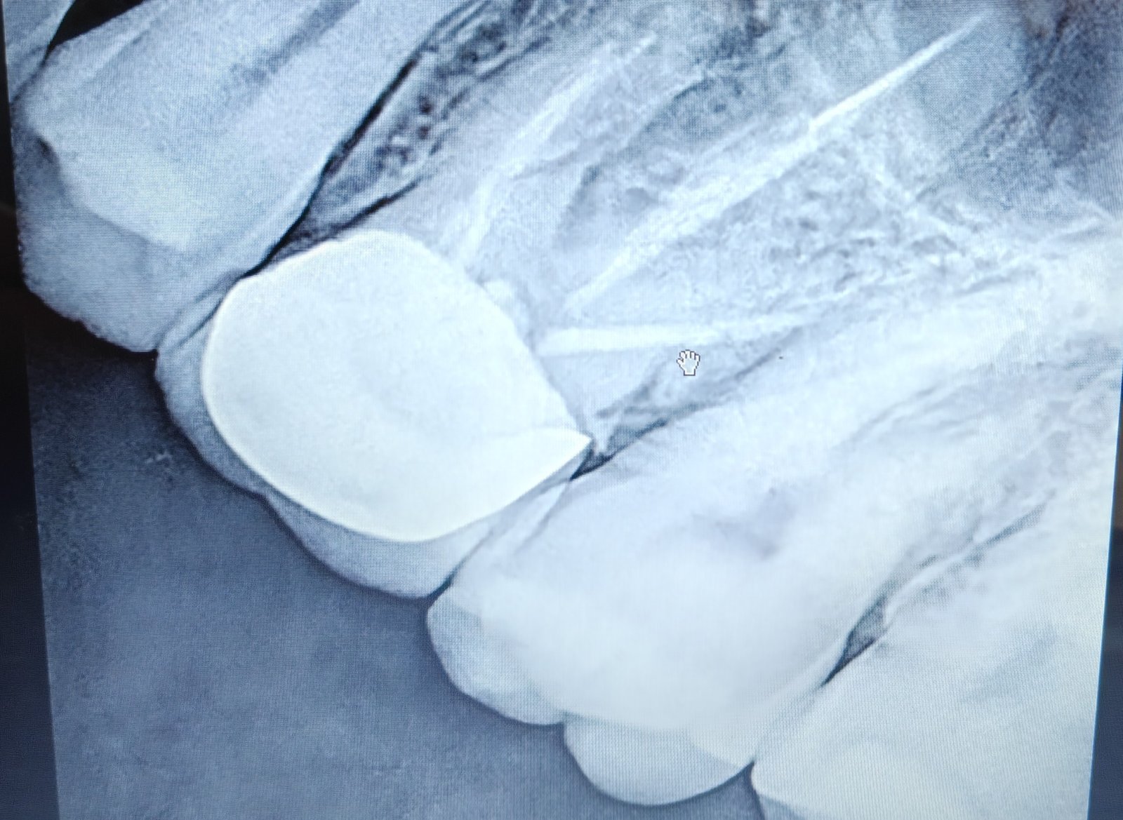
Retrieving a separated file from the palatal root of an upper molar is sometimes possible. It becomes unpredictable when the fragment lies deep or beyond a curve, because removing it can require cutting away so much root dentine that the tooth is left weaker than before. Combined with the crown fracture, the prognosis was poor. In that situation the most honest advice is to remove the tooth and plan a replacement that protects the bone, rather than keep repairing something that will fail.
Above the roots of upper molars lies the maxillary sinus, an air-filled cavity. After a molar is lost, the bone between the socket and the sinus floor often shrinks. If an implant of adequate length is placed without preparation, it can push through into the sinus. The usual answer is a sinus lift, in which the sinus membrane is gently raised and the space is filled with bone graft.
| Approach | How it works | Best suited to |
|---|---|---|
| Direct (lateral window) sinus lift | A window is opened in the side wall of the bone to lift the membrane. Usually a separate surgery with a longer healing period. | Very little remaining bone height |
| Indirect (crestal) sinus lift | The membrane is elevated through the implant site itself, from below, with the graft placed through the same opening. | Moderate bone height, where it can be combined with implant placement |
In this case, assessment of the available bone allowed an indirect sinus lift, which spared the patient a second, more invasive surgery.
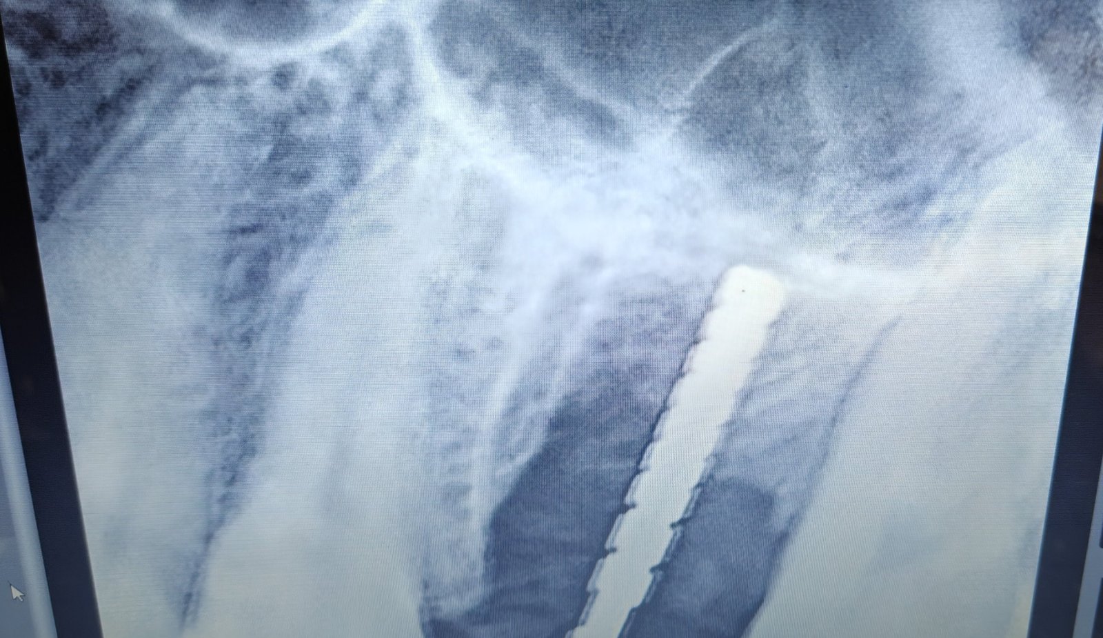
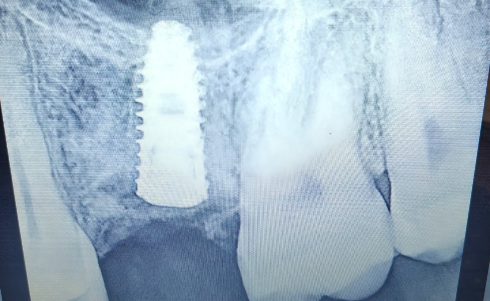
Healing and next steps. After placement, the implant needs time to fuse with the bone before a permanent crown is attached. Individual healing times vary, and we confirm readiness with clinical and radiographic review.
MDS in Maxillofacial Prosthodontics & Oral Implantology (MCODS, Manipal), with 16+ years of specialist experience.
Full profile →
MDS in Oral & Maxillofacial Surgery (Manipal), with 16+ years of specialist experience.
Full profile →Complex implant cases succeed through teamwork, precise planning and respect for anatomy. Our approach at Dental Arch, Gurgaon is to explain every option honestly, including when a tooth should and should not be saved.
With local anaesthesia, the procedure itself is not painful. Mild swelling or tenderness afterwards is common and is managed with medication and care instructions.
In suitable cases, yes. It depends on the condition of the socket, the amount of bone, and the absence of active infection. Not every tooth qualifies, which is why assessment comes first.
Many separated fragments are managed without harm. When the tooth is also structurally compromised, as here, extraction and replacement may be the most predictable path.
Book a consultation with Dr. Anish Goyal and Dr. Neha Sharma at Dental Arch, Gurgaon. Bring your X-rays, and we will explain your options clearly.
Patient details have been anonymised and published with the patient's consent. Outcomes vary between individuals; this report describes a single case and is for information, not a substitute for a personal consultation.
Dental Arch Gurgaon
Lower Ground Floor-105, Sector-28, Gurgaon, Haryana. Near Golf Course Road.